


 3D scanner / tomographic / with magnetic particle imaging (MPI)
MPI PreClinical
Vnation JSC

Contact us for advice solutions and equipment provider
Products relative
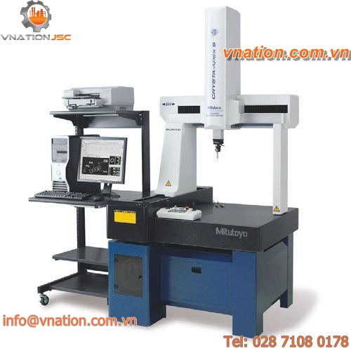
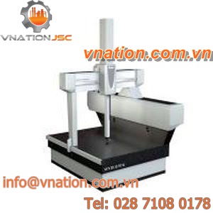
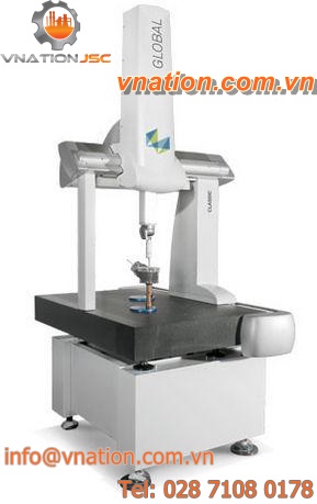
Coordinate measuring machines: bridge type CMMs, horizontal arm CMMs
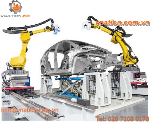
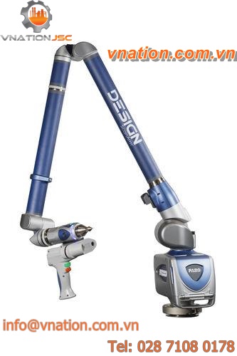
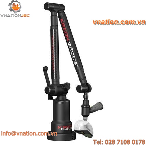
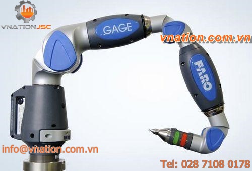
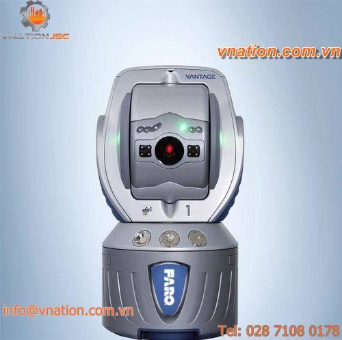
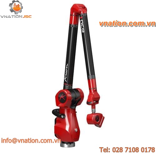
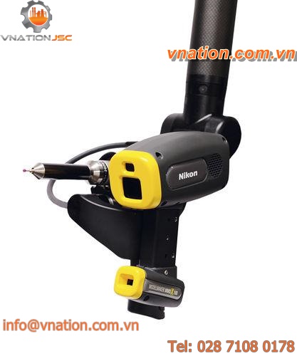
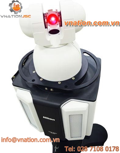
3D measuring arms, laser trackers

Form and surface measuring instruments